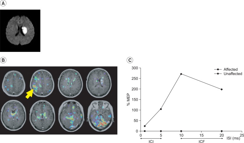
Fig. 2.
A patient in a fair recovery group (Patient 3). (A) Brain MRI showed an acute infarct in the left corona radiata. (B) During the motor task, activation occurred in the contralateral motor cortex, the cerebellum, the perilesional area, bilateral frontal cortices and thalamus. (C) No MEP was observed in the affected APB, but normal ICI and ICF patterns were observed in the unaffected side. ICI: Intracortical inhibition, ICF: Intracortical facilitation, ISI: Interstimulus interval, ms: Milliseconds.

