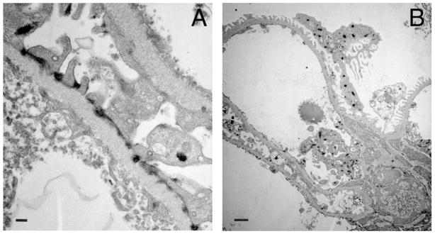
Figure 1. Expression of VEGFR2 in vivo.
Transmission electron microscopy from Flk-1-LacZ 6 kidney showing LacZ deposits en lieu of VEGFR2 localized to A) foot processes near slit-diaphragm, GBM and fenestrated endothelium; B) podocytes and endothelial cell. Scale bars= 200nm (A) and 1 μm (B). Sample processed as reported.26

