FIGURE 3.
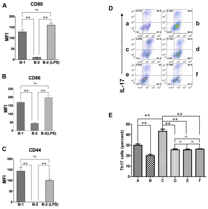
Activated B-2 cells stimulate Th17 cell differentiation like B-1 cells. (A–C) Peritoneal B-1 cells and splenic B-2 cells were evaluated for expression of CD80 (A), CD86 (B), and CD44 (C) by immunofluorescent staining and flow cytometry. Splenic B-2 cells were sort-purified and stimulated by 10 μg/mL LPS and then similarly evaluated. Mean values are shown along with lines indicating SEMs (n = 3). (D,E) Sort-purified BALB/c peritoneal B-1 cells (a,b) and splenic B-2 cells previously stimulated by 10 μg/mL LPS (c–f) were co-cultured at a 1:2 ratio with magnetic bead selected CD4+ T cells from CD57BL/6 mice in Th17 polarizing conditions in medium alone (a,c) or in the presence of anti-CD44 antibody (d), anti-CD80 plus anti-CD86 antibody (e) or anti-osteopontin antibody (b,f). After 5 days expression of intracellular IL-17 was assessed by flow cytometry. Representative results are shown in (D) and mean values (with horizontal lines indicating SEM) for five independent experiments are shown in (E). In (A,B,C,E), t-test was performed to determine whether differences were significant. **p < 0.01; *p < 0.05.

