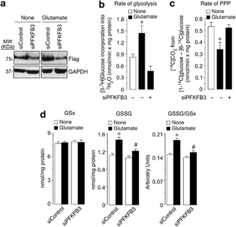
Figure 3.
Glutamate stimulates PFKFB3-dependent increase in glycolysis, a decrease in PPP and promotes glutathione oxidation in neurons. (a) Incubation of GFP-PFKFB3-expressing neurons with glutamate (100 μM/15 min) induced, 6 h after treatment, PFKFB3 accumulation in control, siRNA-treated neurons (siControl), as revealed by an anti-GFP (Flag) antibody; transfection of neurons with an siPFKFB3 efficiently reduced PFKFB3 protein and prevented glutamate-induced PFKFB3 accumulation. (b) Incubation of neurons with glutamate (100 μM/15 min) increased, after 6 h, the rate of glycolysis, as assessed by the determination of [3-3H]glucose incorporation into 3H2O; this effect was abolished by preventing PFKFB3 accumulation in neurons previously transfected with siPFKFB3. (c) Glutamate treatment decreased, after 6 h, the rate of the PPP, as assessed by the determination of the difference between 14CO2 produced by [1-14C]glucose and that of [6-14C]glucose; this effect was abolished by siPFKFB3. (d) Glutamate treatment did not change GSx (left panel), but it increased GSSG (middle panel) and the oxidized glutathione redox status (GSSG/GSx; right panel); these effects were partially prevented by siPFKFB3. *P<0.05 versus none; #P<0.05 versus the corresponding siControl (ANOVA)

