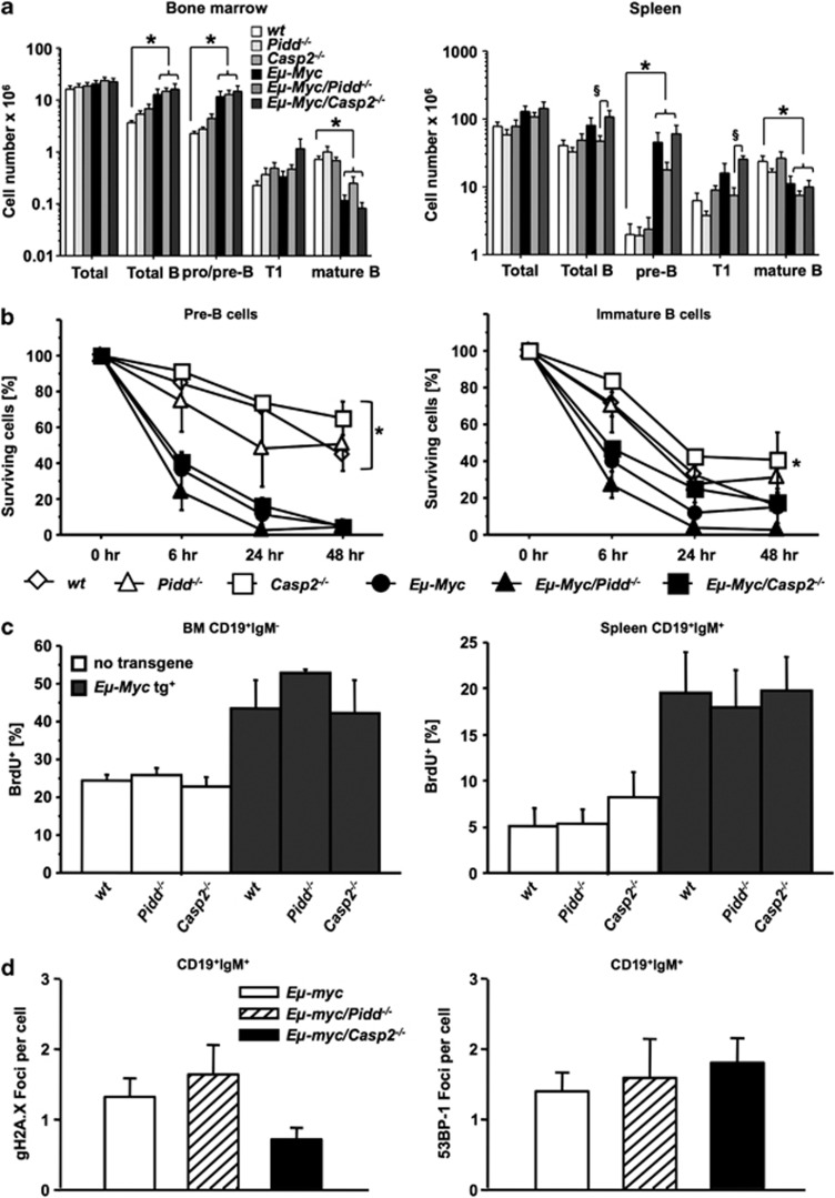
Figure 4.
Normal cell death and proliferation rates in PIDDosome-defective premalignant B cells. (a) Distribution of B-cell subsets in bone marrow, or spleens of mice of the indicated genotypes. Single-cell suspensions derived from premalignant animals 5 weeks of age were counted, stained for different B-cell markers and analyzed by flow cytometry. Data are represented as means of n>4 animals/genotype ±S.E.M. *P<0.05 compared with Eμ-Myc controls, §P<0.05 compared with Eμ-Myc/Casp2−/− mice. (b) Sorted pre-B cells from the bone marrow (CD19+IgM−CD43−), immature B cells (IgM+IgDlow) from the spleens of mice of the indicated genotypes were cultured for 0, 6, 24 and 48 h cells and cell survival in CD19+ B cells was monitored by Annexin V/PI staining and flow cytometric analysis. Data points represent mean values±S.E.M. from n>3 cell mice/genotypes. *P<0.05 all Eμ-myc versus all non-transgenic genotypes (ANOVA and Student–Newman–Keuls test). (c) BrdU incorporation was determined in CD19+IgM− pro/pre-B cells isolated from bone marrow (left) and mature CD19+IgM+ B cells isolated from spleen (right) from indicated genotypes (5 weeks old). Data are mean values±S.E.M from n>4 mice/genotypes. (d) Mature B cells (CD19+IgM+), sorted from spleens derived from premalignant mice of the indicated genotypes, were stained for γH2AX or 53BP-1 and foci/cells is depicted as bar graphs of mean values±S.E.M. of n>3 preparations from individual animals

