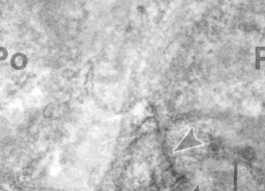
Figure 2.
Post-fixation immunoperoxidase electron microscopy of developing mouse glomerular capillary wall using anti-collagen α3α4α5(IV) IgG. Peroxidase reaction product appears within the GBM and intracellular biosynthetic organelles (arrowheads) of podocytes (Po). Labeling is absent in the glomerular endothelium (En). Reprinted with permission.34

