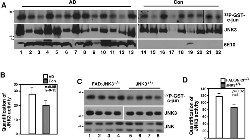
Figure 3. JNK3 activity is increased in human AD and FAD cases.
(A) JNK3 activity is increased in human AD cases compared to age-matched controls. The lysates from human frontal cortices were subjected to JNK3 immunoprecipitation/kinase assays using GST-c-jun as a substrate. As a control, JNK3 and 6E10 Western blots are also shown.
(B) Quantification of 32P-GST-c-jun in (A). The data are represented as means ± SEM.
(C) JNK3 activity is higher in FAD mice compared to normal mice at 5-6 months. Lanes 1 to 4 represent four different FAD:JNK3+/+ mice, and lanes 5 to 8, JNK3+/+ mice.
(D) Quantification of 32P-GST-c-jun in (C). The data are represented as means ± SEM.

