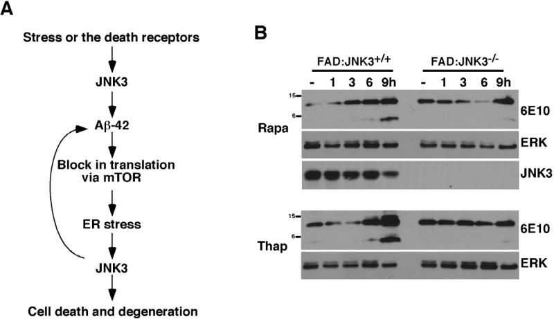
Figure 8. Translational block exacerbates APP processing in JNK3-dependent manner.
(A) A model of how JNK3 activation exacerbates the development of AD pathology. We hypothesize that JNK3 is initially activated by death receptors and/or under certain stress conditions. Once activated, JNK3 phosphorylates APP at T668P, facilitating APP processing by enhancing its internalization, which results in greater Aβ42 production. Aβ42 then blocks translation, leading to UPR, which activates JNK3. Activated JNK3 will target APP again in a positive feedback loop, thereby perpetuating a cycle that exacerbates Aβ42 production.
(B) Both Thapsigargin and Rapamycin increase Aβ42 production in a JNK3-dependent manner. Organotypic brain slices from FAD:JNK3+/+ and FAD:JNK3-/- were treated with the vehicle, 0.5 μM Thapsigargin, and 10 nM Rapamycin for the indicated amount of time and the resulting whole cell lysates were analyzed for 6E10 Western. In order to facilitate detection of CTF and Aβ peptides, the slices were also treated with 10 μM MG132 at the same time. Note that both CTF and Aβ42 levels were increased in FAD:JNK3+/+ cultures upon treatments compared to those in the vehicle control. This increase was significantly attenuated in FAD:JNK-/- cultures.

