
Figure 3.
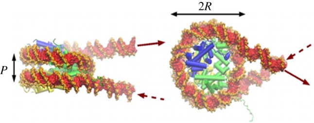
Structure of the nucleosome. In green, the histone tetramer (H3-H4)2, in blue and yellow, the two histone dimers (H2A-H2B). The figure has been obtained starting from the crystallographic structure [31], to which were added two short entering and exiting DNA segments (courtesy of Richard Lavery).