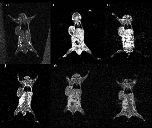
Figure 3.

Coronal slices under T1-weighted tumor imaging pre- and post-injection of Gd-DTPA. (a) Pre-injection scanning; (b) post-injection; (c) post-12 h injection; (d) 24 h time-point post-injection; (e) 48 h time-point post-injection; (f) 72 h time-point post-injection.
