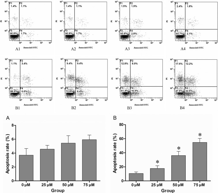
Figure 1.
(A1–4) CNE-2 cells were treated with different concentrations of DAPT (0, 25, 50 and 75 μM), and the apoptosis of CNE-2 cells was assessed by FACS after 48 h. (B1–4) CNE-2 cells were previously treated with various concentrations of DAPT (0, 25, 50 and 75 μM) before 24 h, and then treated with cisplatin at the same final concentration of 10 μM. The apoptosis of CNE-2 cells was detected by FACS after 48 h of treatment with cisplatin. The control group (0 μM DAPT) was treated with conventional medium containing the mean volume of DMSO. No significant apoptosis was noted only with DAPT treatment when compared to the control group (P>0.01), while pre-treatment with DAPT enhanced the effect of cisplatin in a dose-dependent manner. There was a marked difference compared to the control group treated with cisplatin and DMSO (P<0.01). A1–4 and B1–4 are respresentative of one of three independent experiments that yielded similar results. Values in histograms (A) and (B) are the means ± SD. Proportion of non-apoptotic cells (F3, Annexin V-FITC−/PI−), early apoptotic cells (F4, Annexin V-FITC+/PI−), late apoptotic/necrotic cells (F2, Annexin V-FITC+/PI+) and cell debris or dead cells (F1). *P<0.01 compared to that of 0 μM DAPT group by T-test.

