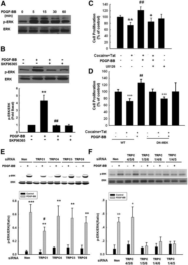
Figure 5.
TRPC channels are involved in PDGF-BB-induced activation of the ERK pathway. A, PDGF-BB induced time-dependent phosphorylation of ERK. B, Representative immunoblot of NPCs exposed to PDGF-BB in the presence of the TRPC blocker SKF96365 (20 μm) was monitored for ERK activation. Densitometric analysis of p-ERK/ERK from four individual experiments. **p < 0.01 versus control group; ##p < 0.01 versus PDGF-BB group. C, Pretreatment of NPCs with the MEK inhibitor U0126 (10 μm) for 1 h significantly attenuated the effect of PDGF-BB on the restoration of Tat–cocaine-mediated impairment of proliferation. Data are presented as mean ± SD of four individual experiments. **p < 0.01 versus control group; ##p < 0.01 versus Tat–cocaine-treated group; +p < 0.05 versus both PDGF-BB-treated and Tat–cocaine-treated groups. D, PDGF-BB failed to restore impaired proliferation induced by Tat–cocaine in the presence of DN MEK but not in the wild-type group. Data are presented as mean ± SD of four individual experiments. ***p < 0.001 versus control group; ##p < 0.01 versus Tat–cocaine-treated group. E, Representative immunoblot of PDGF-BB-mediated ERK phosphorylation in NPCs transfected with TRPC1, TRPC4, TRPC5, or TRPC6 siRNAs (top), followed by densitometric analyses from three individual experiments (bottom). F, NPCs were knocked down for three TRPC channels with the expression of only one channel and were then monitored for PDGF-BB-mediated ERK phosphorylation (top), followed by densitometric analyses from three individual experiments (bottom). *p < 0.05, **p < 0.01, ***p < 0.001 versus control group; #p < 0.01 versus PDGF-BB-treated cells transfected with nonsense (Non) siRNA group.

