Figure 9.
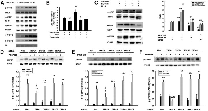
TRPC channels are involved in PDGF-BB-induced activation of mTOR/4E-BP–p70S6K pathways. A, PDGF-BB induced time-dependent phosphorylation of mTOR/4E-BP–p70S6K/S6. B, Pretreatment of NPCs with the mTOR inhibitor rapamycin (1 μm) for 1 h significantly attenuated the effect of PDGF-BB in restoring impaired proliferation induced by Tat–cocaine. Data are presented as mean ± SD of four individual experiments. **p < 0.01 versus control group; ##p < 0.01 versus Tat–cocaine-treated group; +p < 0.05 versus both PDGF-BB-treated and Tat–cocaine-treated groups. C, Representative immunoblot of NPCs exposed to PDGF-BB in the presence of the TRPC blocker SKF96365 (20 μm) or the MEK inhibitor U0126 (10 μm) monitored for mTOR/4E-BP–p70S6K activation (left). Densitometric analysis of mTOR/4E-BP and p70S6K from four individual experiments (right). **p < 0.01, ***p < 0.001 versus control group; #p < 0.05, ##p < 0.01, ###p < 0.001 versus PDGF-BB group. Representative immunoblot of PDGF-mediated mTOR (D), 4E-BP (E), and p70S6K (F) phosphorylation in NPCs transfected with TRPC1, TRPC4, TRPC5, or TRPC6 siRNAs (top). Densitometric analysis of PDGF-BB-mediated phosphorylation of mTOR/4E-BP–p70S6K6 in NPCs transfected with TRPC1, TRPC4, TRPC5, or TRPC6 from three individual experiments (bottom). *p < 0.05, **p < 0.01, ***p < 0.001 versus control group; #p < 0.05, ###p < 0.001 versus PDGF-BB-treated cells transfected with nonsense (Non) siRNA group.

