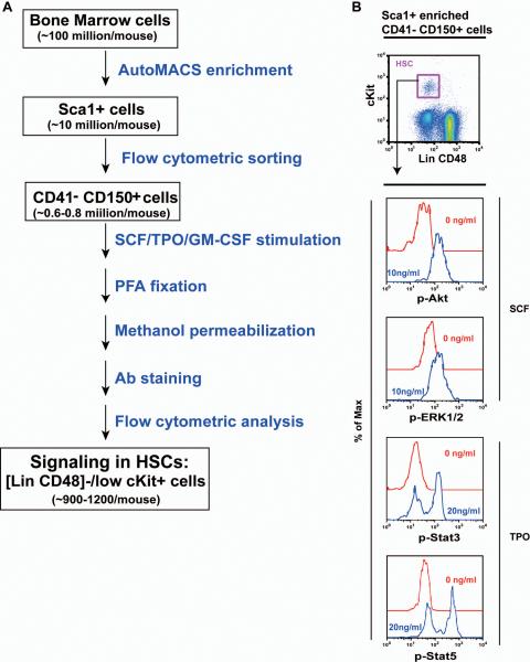
Figure 2. HSCs are highly responsive to SCF and TPO stimulation.
(A) Flow chart of the developed “HSC phospho-flow” method. (B) Sca1+ enriched, CD150+ CD41− bone marrow cells were serum- and cytokine-starved for 30 minutes and stimulated with 10 ng /ml of SCF or 20 ng/ml of TPO for 10 minutes at 37°C. Levels of phosphorylated ERK1/2, Akt, Stat3, and Stat5 were measured using phospho-specific flow cytometry. HSCs (defined as [Lin CD48]−/low cKit+ cells) were gated for data analysis. Representative gating strategy and plots of phosphorylated signaling proteins are shown (n= 6).

