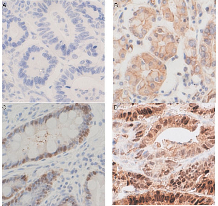
Figure 1.
Representative immunohistochemical maspin staining in gastric cancer. Tumor cells with (A) negative nuclear and cytoplasmic staining, (B) negative nuclear and weakly positive cytoplasmic staining, (C) weakly positive nuclear and negative cytoplasmic staining, (D) positive nuclear and cytoplasmic staining.

