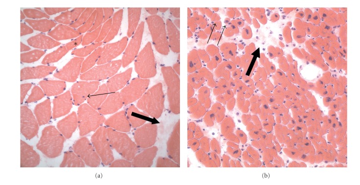
Figure 1.
Histology (hematoxylin-eosin staining) of pectoralis major muscle (a) and heart muscle (b). In the pectorals major, mild myopathic changes with hypotrophic fibers (asterisk), some internal nuclei (small arrow), and an increase of fibrous tissue (large arrow) are seen. In the myocardium, there are some atrophic cardiomyocytes (small arrows) and a mild increase in connective tissue.

