
Figure 1.
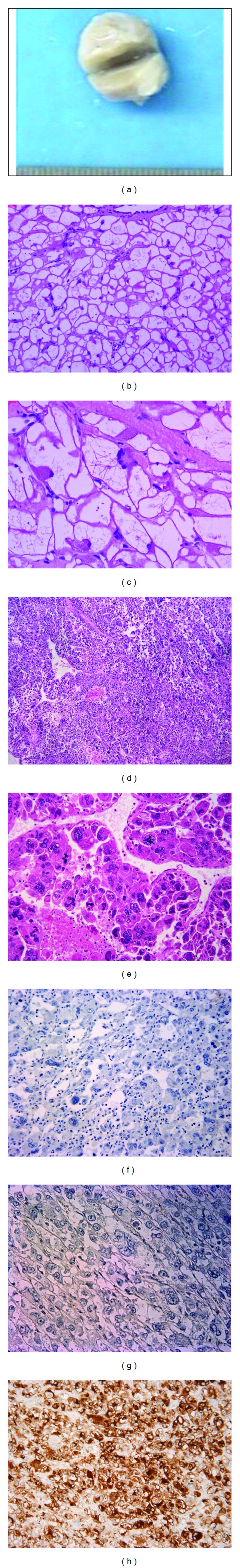
Epithelioid PEComas in hearts. Figures 1(a)–1(c): Case 1. (a) gross picture for case 1. (b)-(c) H&E for case 1 ((b) ×100; (c) ×200); Figures 1(d)–1(h). Case 2: metastasis. (d) tumor with necrosis, H&E, ×50; (e) multinuclear giant cells with nuclear atypia and atypical mitosis, H&E, ×200; (f) case 2, negative cytokeratin (AE1/3) stain, ×200; (g) case 2, negative S100 stain, ×200; (h) case 2, diffusely positive HMB-45 stain, ×200.