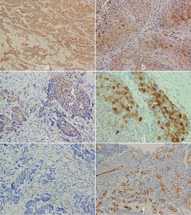
Figure 1.
The representative immunohistochemical staining of angiogenic markers in upper tract urothelial carcinoma, with strong cytoplasmic VEGF activity in high grade (A); altered cytoplasmic expression of VEGFR1 and (B); VEGFR2 in invasive tumors with (C), nuclear and cytoplasmic staining of HIF 1α (D). Immunostaining of endothelial cells for CD31 and (E). active areas of angiogenesis detected with CD34 in urothelial cancer (F) (original magnification: x400).

