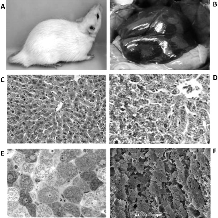
Figure 2.
Images of a control rat. (A) External features of a rat showing normal bright colored fur and healthy-appearing tail. (B) Gross morphology of healthy liver with normal reddish-brown color. (C) H&E section of liver tissue as in panel B; magnification, ×240. (D) Toluidine blue section of liver tissue as in panel B; magnification, ×300. (E) Masson’s trichrome staining of liver tissue; magnification, ×400. (F) SEM image showing fractured surface taken from within the liver parenchyma; magnification, ×1,000.

