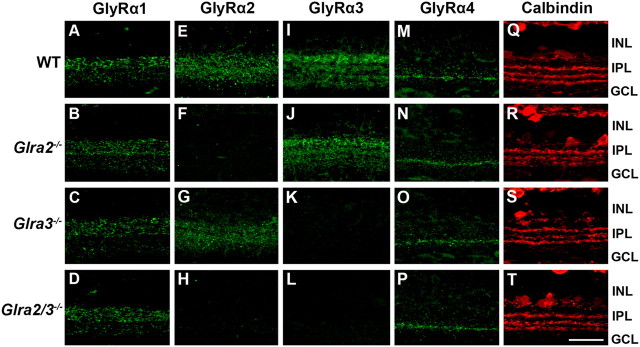
Figure 2.
GlyRα subunit expression across WT, Glra2−/−, Glra3−/− and Glra2/3−/− retinas. A–T, Representative confocal images of transverse retinal sections (25 μm) reacted with one of four GlyRα subunit-specific antibodies. INL, Inner nuclear layer; IPL, inner plexiform layer; GCL, ganglion cell layer. Scale bar, 20 μm. GlyRα1-immunoreactive puncta are prominent in the outer strata of the IPL across all genotypes (A–D). Representative confocal images of GlyRα2-immunoreactive puncta are diffuse across all IPL strata in WT and Glra3−/− retinas (E, G) and are absent in Glra2−/− and Glra2/3−/− retina (F, H). GlyRα3-immunoreactive puncta label four bands in the IPL of WT and Glra2−/− retinas (I, J) and are absent in the Glra3−/− and Glra2/3−/− retina (K, L). GlyRα4-immunoreactive puncta are localized to a distinct band within the IPL in all genotypes (M–P). Retinas of all genotypes were reacted with an antibody to calbindin (red) and illustrate that IPL sublamination patterns are similar across genotype (Q–T).

