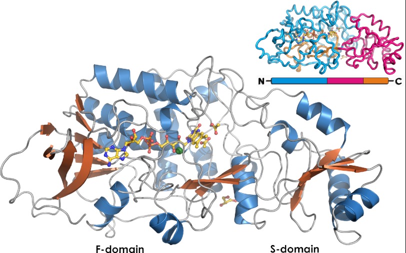
FIGURE 4.
Overview of the Δ4-(5α)-KSTD structure. FAD, glycerol, and the two acetate molecules bound in the active site are shown in yellow. The Cl− ion is shown in green. The inset shows the domain organization of Δ4-(5α)-KSTD. Domain F is shown in blue (F1) and orange (F2), and the S domain is in pink. Domain S is an insert into domain F, and the active site is located on the interface between the two domains.

