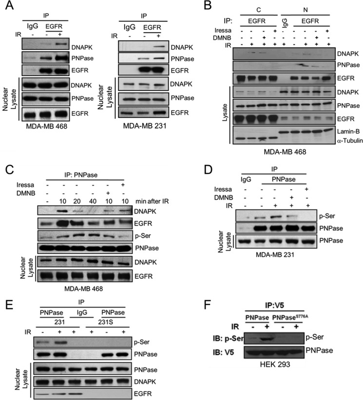
FIGURE 3.
EGFR regulates DNAPK-mediated serine phosphorylation of PNPase at Ser-776 upon ionizing radiation. A, MDA-MB 468 and MDA-MB 231 cells were irradiated with or without 4 Gy IR. After 10 min, nuclear lysates were harvested and individually immunoprecipitated with antibodies against IgG or PNPase, followed by SDS-PAGE separation and IB (upper panel). The endogenous expression levels of the indicated protein were examined by IB (lower panel). B, MDA-MB 468 cells were irradiated with or without 4 Gy IR in the presence or absence 10 μm DMNB or 10 μm Iressa. After indicated treatment, cytosolic (C) and nuclear (N) lysates were harvested and individually immunoprecipitated with antibody against IgG or EGFR, followed by SDS-PAGE separation and IB (upper panel). The endogenous levels of DNAPK and PNPase are shown in the lower panel. C, same as B, at the indicated time interval, The nuclear lysates of MDA-MB 468 cells were harvested and individually IP with antibody against IgG or PNPase, followed by SDS-PAGE separation and IB (upper panel). The endogenous levels of DNAPK and EGFR were shown in the lower panel. D, MDA-MB 231 cells were irradiated with or without 4 Gy IR in the presence or absence 10 μm DMNB or 10 μm Iressa. After 10 min, nuclear lysates were harvested and individually immunoprecipitated with antibody against IgG or PNPase, followed by IB for phospho-serine (p-Ser) (upper panel). The endogenous expression level of PNPase was shown in the lower panel. E, MDA-MB 231 radio-resistant (231) and radio-sensitive (231S) cells were irradiated with or without 4 Gy IR. After 10 min, nuclear lysates were harvested and individually immunoprecipitated with antibody against IgG or PNPase, followed by IB for phospho-serine (p-Ser) (upper panel). The endogenous expression level of PNPase was shown in the lower panel. F, HEK-293 cells were transfected with plasmid harboring V5-tagged PNPase or PNPaseS776A. These transfectants were exposed to 0 or 4 Gy IR. After 10 min, cell lysate was extracted and immunoprecipitated with anti-V5 antibody, followed by IB for p-Ser (upper panel) or V5 (lower panel).

