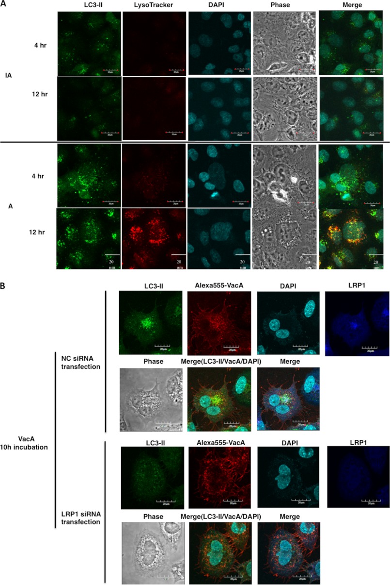
FIGURE 4.
VacA induced formation of autophagic vacuoles in AZ-521 cells via LRP1. A, VacA-induced formation of autophagosomes and autolysosomes in AZ-521 cells. AZ-521 cells were incubated with 120 nm heat-inactivated (IA) or wild-type VacA (A) for the indicated time points and fixed for immunofluorescence staining with LC3B (green) antibodies as described under “Experimental Procedures.” The acidic autophagolysosomes were stained by LysoTracker, as described under “Experimental Procedures.” A merged picture shows co-localization in AZ-521 cells. The nuclei were stained with DAPI. Bars represent 20 μm. Experiments were repeated two times with similar results. B, induction of autophagy by VacA in an LRP1-dependent manner. The indicated siRNA-transfected AZ-521 cells were incubated with 120 nm Alexa 555-labeled VacA (red) for 10 h at 37 °C and fixed for immunofluorescence staining with anti-LC3B (green) or anti-LRP1 (blue) antibodies as described under “Experimental Procedures.” A merged picture shows co-localization in AZ-521 cells. The nuclei were stained with DAPI. Bars represent 20 μm. Experiments were repeated two times with similar results.

