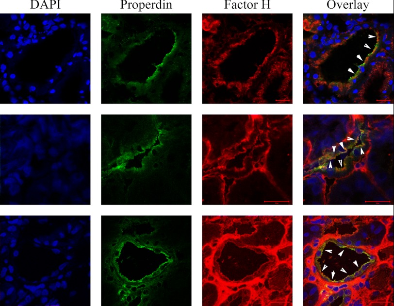
FIGURE 2.
Factor H and properdin colocalization on luminal side of tubuli. Factor H and properdin are both present on the luminal side of tubular cells in adriamycin-induced nephrosis. The nuclei are shown in blue, properdin is in green, and factor H is in red. The white arrowheads show colocalization areas of factor H and properdin on the luminal side of tubular cells. The scale bars represent 20 μm in all images.

