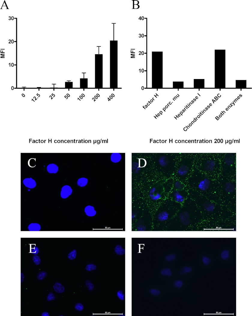
FIGURE 3.
HS-dependent binding of factor H to HK2 cells. A, factor H binds to HK2 cells in a dose-dependent manner in flow cytometry analysis; this graph shows the result of one (performed in duplicate) of four experiments. The results are expressed as mean fluorescence intensity (MFI) ± S.E. B, in a representative FACS experiment of three, HK-2 cells were incubated either with heparitinase I, chondroitinase ABC, or both heparitinase I and chondroitinase ABC before factor H incubation (100 μg/ml). Factor H binding was detected on the cells by FACS staining. The results are expressed as mean fluorescence intensity. C–F, immunofluorescent staining of HK2 cells. C, cultured HK2 cells do not express factor H. D, exogenous factor H (150 μg/ml) binds to HK2 cells. E, preincubation of exogenous factor H (150 μg/ml) with heparin (300 μg/ml) impairs the binding of factor H to these cells. F, heparitinase I pretreatment of HK2 cells abolish factor H binding to the cells. The scale bars represent 50 μm.

