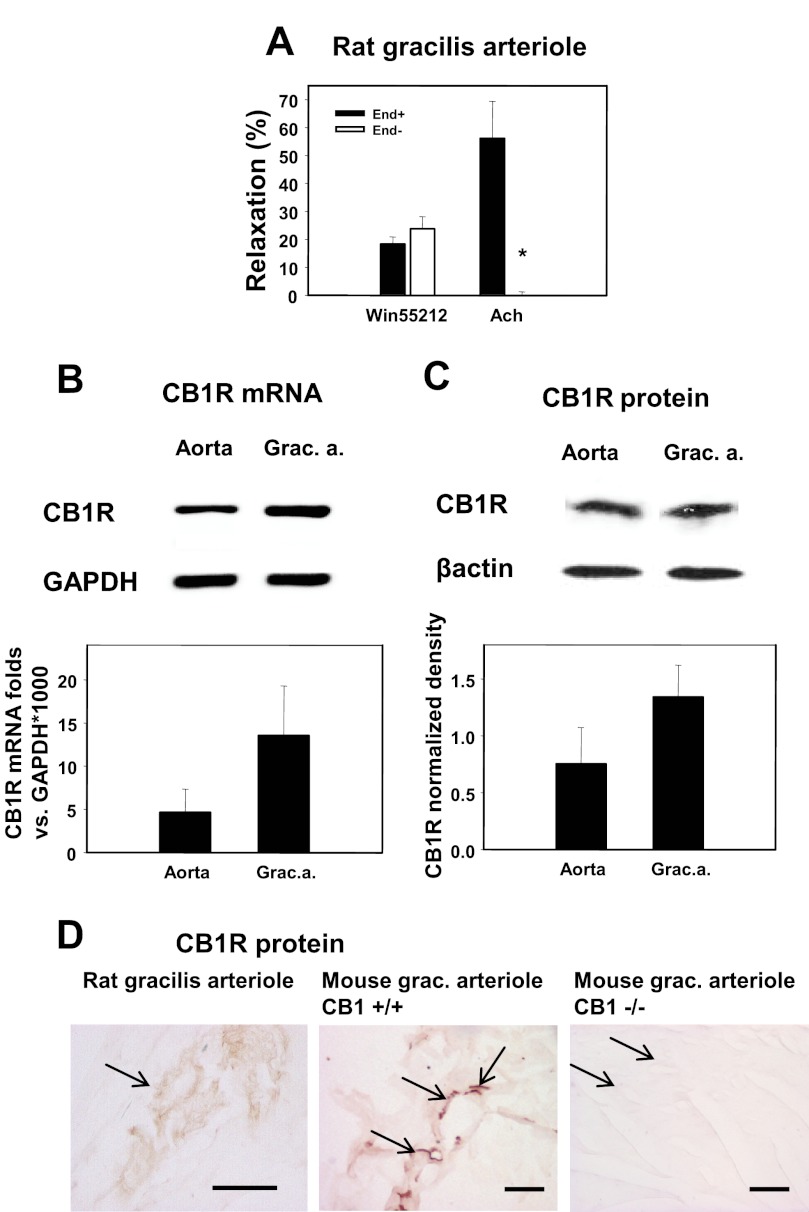
FIGURE 1.
CB1R receptor expression and function in rat skeletal muscle arterioles. A, relaxations induced in rat gracilis arteriole segments by the CB1R agonist WIN 55212 (1 μm) and by the endothelial relaxant Ach (10 μm) as well as the effects of de-endothelization (5–5 segments) are shown. Values were calculated as percent change of diameter compared with control. Mean ± S.E. values are shown. The asterisk indicates significant change of agonist-induced tone in response to inhibitor treatment (p < 0.05). B, shown is gel electrophoresis of the mRNA amplicons and relative quantification of mRNA expression. Messenger RNA expression of CB1R (Cnr1 gene) was normalized to glyceraldehyde-3-phosphate dehydrogenase (Gapdh, n = 4). Grac. a., gracilis arterioles. C, shown is Western blot detection of CB1R protein from tissue homogenates. Expression of CB1R protein in rat aorta and gracilis arterioles was quantitatively detected by densitometry (n = 4) of HRP-induced fluorescence. D, immunohistochemical localization of the CB1R protein (arrows) in rat gracilis arterioles and in mouse gracilis vessels is shown. No staining was detected in CB1R knock-out (−/−) mice. Left bar, 50 μm. Bars in middle and right, 100 μm.

