
FIGURE 4.
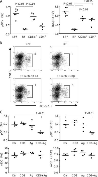
Role of CD8+ T cells in the pDC deficiency of RF mice. A, Low-density cells were prepared from spleen of SPF WT, RF WT, RF CD8α−/−, and RF CD4−/− mice. Cells were stained for pDC as in Fig. 1. pDC frequency (percentage of low-density cells) and absolute numbers in spleen are tabulated; p values were obtained using Student's t test for group comparisons. B, RF mice were treated with anti-NK1.1 and anti-CD8β to deplete NK cells or CD8αβT cells; irrelevant isotype treatment was used as a negative control. Mice received Ab injection twice and were sacrificed 1 wk later after the final injection. Low-density splenocytes were isolated and stained for flow analysis; numbers indicate percentage of total low-density cells. Data are representative of three mice per group. C, SPF mice were injected i.v. with CD8+ T cells from RF mice and injected i.p. with lumenal lysate prepared from RF mice (Ag) or saline (Ctr). Mice were sacrificed 4days later and the frequency (percentage of low-density cells) and absolute numbers of splenic pDC and mDC were determined. Values of p were obtained using Student's t test comparing each group with control mice. Nonsignificant p values (p ≥ 0.05) are not listed.