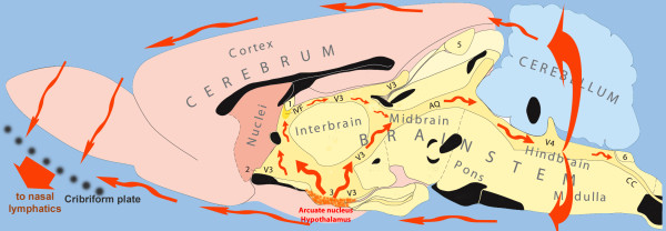
Figure 2.
Diagram showing the flow of CSF in the volume transmission of β-endorphin. The main release site for β-END is the arcuate nucleus of the hypothalamus. The additional hindbrain site is located just ventral to no 6. The flow of the CSF (red arrows) traverses the aqueduct (AQ) to penetrate the mesencephalic periaqueductal gray before reaching the 4th ventricle (V4), and along the ‘vagal-complex’ region. Both regions are important target areas for the flowing β-END. After leaving the ventricular system, the flowing CSF may affect superficial brain regions in the brainstem, hypothalamus and olfactory regions. A considerable part of the CSF and its contents eventually leaves the cranial cavity along the olfactory nerves penetrating the cribriform plate. The telencephalon is indicated in pink colours. The diencephalon (‘interbrain’) is coloured yellow, similar to the brainstem structures and the cerebellum is blue. Black structures show the location of fiber systems which cross the midline. Other symbols: numbers 1–6: circumventricular organs; cc: central canal of spinal cord; IVF: interventricular foramen, connecting the lateral and the 3rd ventricles; V3: 3rd ventricle. (The original figure was kindly provided by L.W. Swanson).

