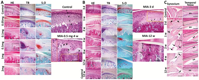
Figure 3. Dose- and time-dependent histopathologic changes in TMJ tissues.
TMJ was sectioned in saggital for HE, TB and S.O staining. A. Dose course (0.05 mg, 0.1 mg, 0.5 mg, 1 mg, 2mg per joint) of MIA 4 weeks post-injection. Black frames are magnified. Condylar cartilage in the controls stained purple blue by TB and red by S.O; (F: fibrous layer; P: proliferative layer; H: hypertrophy layer; C: calcified layer; B: subchondral bone.). Typical OA-like lesion induced by 0.5 mg MIA, including regional loss of chondrocytes (arrow), chondrocyte cluster formation (arrowhead), horizontal cleft (dotted arrow), peripheral chondrocyte proliferation (red frame), and subchondral bone erosion with adjacent bone marrow full of fibroblast-like cells (yellow frame). Lesions staining by TB and S.O was uneven. (Bar = 200 µm) B. Time-dependent changes in the condyle following MIA injection (0.5 mg/joint; 3 days to 12 weeks). Black frames were magnified. After three days, chondrocytes in the anterior and central areas of the cartilage were lightly stained with nuclear condensation (arrowhead). At 12 weeks, thin cartilage (double arrow) and sclerotic subchondral bone (arrowhead) replaced the lesion. (Bar = 200 µm) C. Time-dependent changes in the synovium, disc, and temporal fossa following MIA injection. Time-dependent changes in synovitis (fibrin-like exudates: arrow; proliferative villi of the synovium: dotted arrow), hypo-cellular change and thinning of the disc (arrowhead), and destruction of temporal fossa cartilage (black frame) following MIA induction are shown. (Bar = 300 µm).

