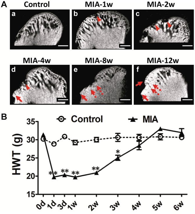
Figure 4. Time-dependent radiographic changes in condylar subchondral bone and nociceptive responses following MIA injection (0.5 mg/joint).
A. Representative images of the condyle by MicroCT scanning with a sagittal section view demonstrated. (a). Control condyle showed intact subchondral bone with a smooth, continuous surface. (b). Regional loss of surface bone (arrowhead) occurred in the frontal bevel of the condyle by 1 week. (c). Multiple erosions of subchondral bone (arrowhead) were observed by 2 weeks. (d). Erosion in the subchondral bone grew deeper and was much more extensive with obvious defects (arrowhead) and osteophyte formation (arrow) by 4 weeks. (e and f). Sclerotic changes (dotted arrow) and osteophytes (arrow) were evident by 8 weeks and 12 weeks. (Bar = 300 µm) B. Changes in animal nociceptive response after MIA injection into TMJ. The HWT was significantly decreased in the first 3 weeks after MIA injection, but gradually recovered to control levels from 4 weeks post-injection. All data were presented as mean ± SEM. (n = 3; **P<0.01; *P<0.05).

