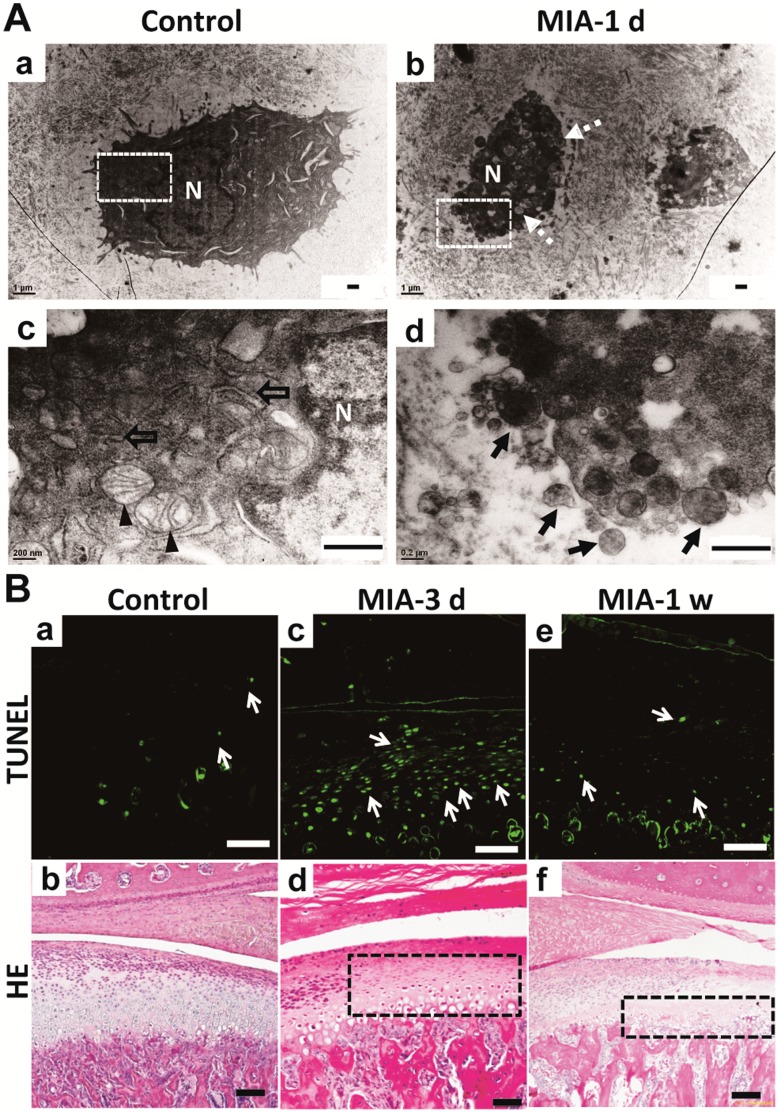
Figure 5. Apoptosis of chondrocytes in condyle after MIA treatment.
A. TEM view of condylar chondrocytes. (a). The chondrocytes in the control group were polygonal. (b). Chondrocytes treated by MIA were shrunken with vacuolar degeneration after 1 day (dotted arrow). (c). Magnified photograph of the white frame in (a). Abundant mitochondria (arrowhead) and endoplasmic reticulum (hollow arrow) were observed around the nuclei (N) in the control chondrocyte. (d). Magnified photograph of the white frame in (b). Apoptotic bodies (black arrow) were observed in the chondrocyte following MIA induction. (Bar = 0.5 µm) B. Comparison of TUNEL assay and HE staining results. (a) There were few apoptotic chondrocytes (arrow) in the control group and the corresponding HE staining shown in (b). (c). Diffuse apoptotic chondrocytes were observed in the region corresponding to the lightly stained area with nuclear condensation of HE staining (black frame in d) at 3 days post-MIA injection. The TUNEL positive chondrocytes almost disappeared (e) due to the extreme loss of chondrocytes as shown by HE staining (black frame in f) at 1 week. (Bar = 80 µm).

