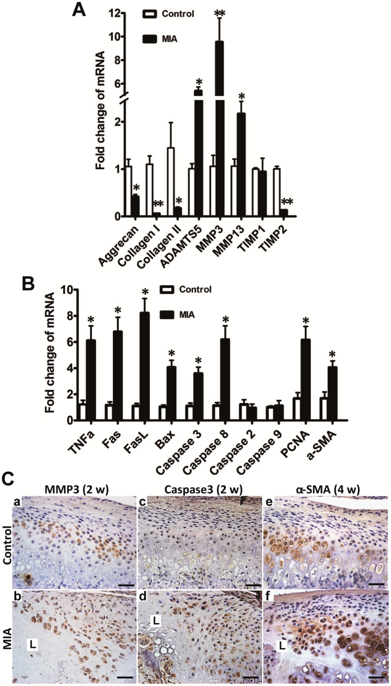
Figure 6. Changes in gene and protein expression in condyle following MIA injection were evaluated by real-time PCR and IHC, respectively.
A. Two weeks after MIA injection, anabolism-associated aggrecan and collagen I and II were downregulated compared with the control group. Catabolism-associated MMP3, MMP13, and ADAMTS5 were upregulated and TIMP2, but not TIMP1, was correspondingly downregulated. B. Two weeks after MIA (0.5 mg) injection, apoptosis-associated genes of the death receptor family, such as, TNFα, Fas, FasL, caspase8, caspase3, and BAX, but not caspase2 and caspase9, were significantly elevated in the MIA injection group; PCNA and α-SMA, representing proliferation and fibrous restoration, respectively, were upregulated (mean ± SEM; n = 6; **P<0.01; *P<0.05). C. There were very few chondrocytes left in the lesion labeled as L 2 weeks after MIA (0.5 mg) injection. MMP3 was mainly expressed in the hypertrophic layer in the control cartilage (a). Diffuse staining of MMP3 was observed in the chondrocytes adjacent to the lesion (L) at 2 weeks (b). Caspase3 was rarely expressed in the control cartilage (c). Enhanced staining of caspase3 was observed in the proliferative and hypertrophic layers adjacent to the lesion (L) at 2 weeks (d). Expression of α-SMA was mainly in the hypertrophic chondrocytes in the control group (e). Stronger staining of α-SMA was observed adjacent to the lesion (L) at 4 weeks (f). (Bar = 40 µm).

