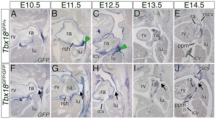
Figure 4. The PPCs in Tbx18-deficient embryos remain open due to the loss of GFP-positive sinuatrial mesenchymal ridges.
(A–J) In situ hybridization analysis of GFP expression on sagittal sections of control and mutant hearts from E10.5 to E14.5. In Tbx18-deficient embryos the mesenchymal ridges are not established (F–J). Stages are as indicated on top and the genotypes on the left side. Black arrows point to the open right PPC. Green arrowheads mark the GFP-positive mesenchymal ridges in control embryos at E11.5 and E12.5. icv, inferior cardinal vein; lu, lung; ppm, pleuropericardial membrane; ra, right atrium; rscv; right superior caval vein; rsh, right sinus horn; rv, right ventricle.

