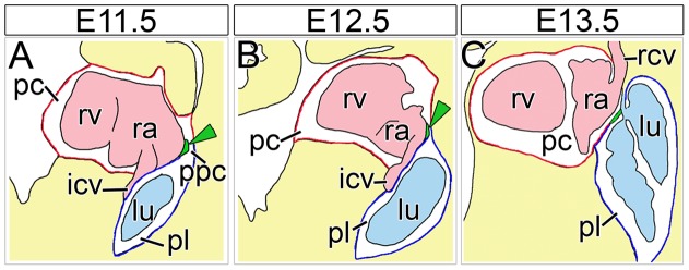
Figure 8. Schematic diagram of PPC closure in mouse development.
(A–C) Scheme of PPC closure by sinuatrial mesenchymal ridges based on sagittal sections through the cardiac venous pole of wildtype embryos from E11.5 to E13.5. Sinuatrial mesenchymal ridges protrude into the PPCs at E11.5 and E12.5 and fuse with the overlying wall of the lung hilus at E13.5 to separate pleural and pericardial cavities. The lumen of the caval veins and the heart is marked in pale pink, the lung and trachea in pale blue, the mesenchymal sinuatrial ridges in green (additionally marked by green arrowheads), the (parietal) pericardium in red, the (parietal) pleura in dark blue and the double-walled PPMs in red and blue. icv; inferior caval vein; lu, lung; pc, pericardial cavity; pl, pleural cavity; ppc, pericardioperitoneal canal; ppm, pleuropericardial membranes; ra, right atrium; rcv, right superior caval vein; rv, right ventricle.

