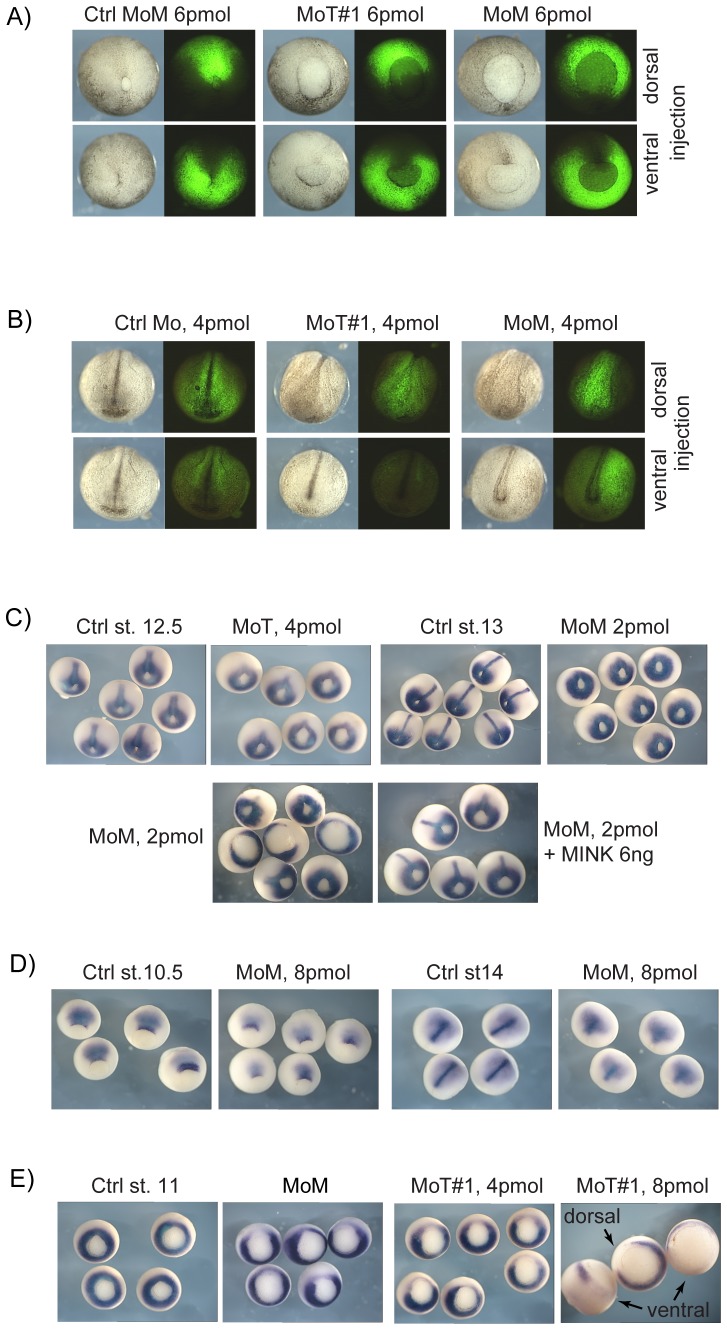
Figure 2. Analyses of xTNIK and xMINK knockdown phenotypes.
A) Posterior views of stage 13 and B) anterior views of stage 19 knockdown embryos. The indicated Morpholinos were injected into the two dorsal or ventral blastomeres of 4 cell embryos together with fluorescein dextran as lineage marker. See Figure S2A and B for additional examples and for dorsal views at stage 19. C) Stage 12.5 knockdown embryos before and after rescue by re-introduction of xMINK were also subjected to in situ hybridization for Xbra mRNA. Extension of the developing notochord is evident in the control embryos and after rescue. D) Similarly stage 10.5 and 14 xMINK knockdown embryos were also hybridized to reveal chordin mRNA. E) Hybridization at stage 11 reveals that knockdown of either xTNIK or xMINK locally suppresses the onset of Xbra expression. Where appropriate, ventral and dorsal sites of knockdown are indicated, otherwise knockdown was targeted to the dorsal blastomeres.

