
Figure 3. Rendering of the whole brain, depicting the major areas (a, b, c) as well as all the 54 delineated structures (d, e, f).
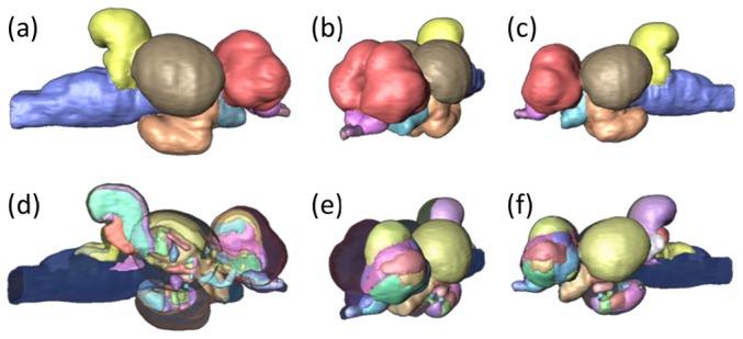
Three different angles are presented to maximize the number of brain regions per image: (a), (d) right view; (b), (e) partial frontal view; (c), (f) left view. In the first row of images it is possible to define six major areas: telencephalon (red), olfactory bulbs (pink) and part of the olfactory tracts (purple), optic tectum (brown) and part of the optic tracts (light blue), diencephalon (orange), cerebellum (yellow) and the brain stem (blue). For a complete list of the small nuclei identified and the color code for the remaining images see Fig. 1.