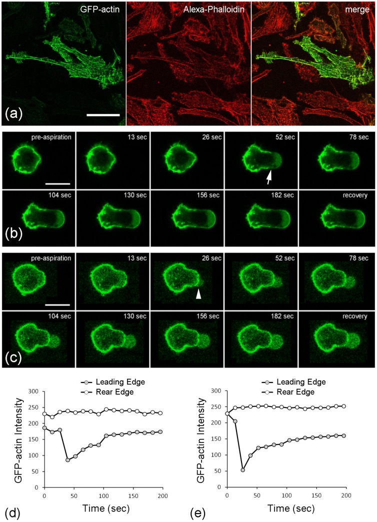
Figure 5. Cell deformation is associated with actin distortion, disassembly and remodelling.
a) Representative confocal images showing characteristic actin organisation in a chondrocyte cultured in monolayer and transfected with GFP-actin (green). F-actin has been co-labelled with alexa-phalloidin (red). Selected images from time series showing GFP-actin distortion and dynamics during micropipette aspiration applied at a) 0.35 and b) 5.48 cmH2O/sec. Arrow indicates breakdown/fluidization of the actin cortex. Arrowhead shows initiation of a membrane bleb. All scale bars represent 10 microns. The corresponding temporal changes in GFP actin intensity at the leading edge (ROI 1) and the rear edge (ROI 2) are shown for a cell aspirated at d) 0.35 and e) 5.48 cmH2O/sec.

