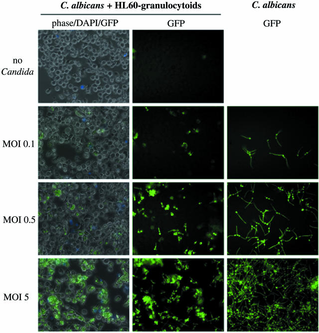
FIG. 3.
HL60 granulocytoid-C. albicans interaction. GFP-expressing C. albicans were cultured either alone (right column) or with HL60 granulocytoids at the indicated MOIs, as described in Materials and Methods. Photographs were taken 1.5 h later at 400× magnification. Each of the panels in the first column is a superimposition of three images: phase contrast, blue fluorescence (to visualize DAPI staining), and green fluorescence (to visualize GFP-C. albicans). Panels in the second column show the images corresponding to the green fluorescence in the first column. Panels in the last column show green fluorescence images to visualize C. albicans cultured alone at densities corresponding to the indicated MOIs.

