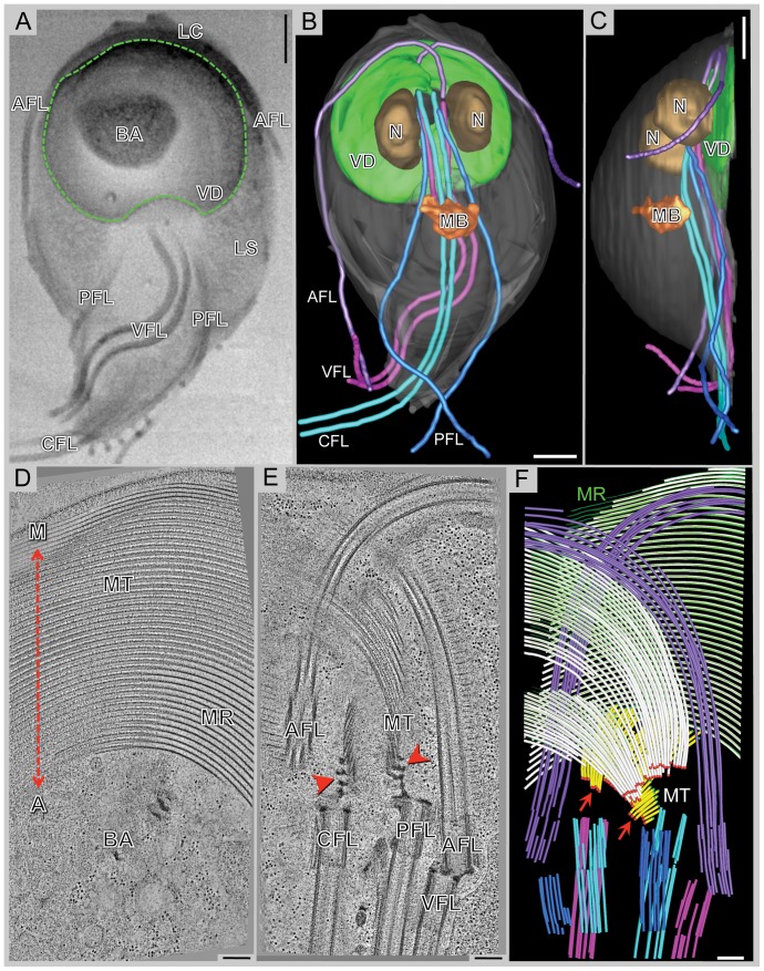
Figure 1. The complex microtubule cytoskeleton of Giardia reconstructed by 3View® and plastic-section tomography.
A) Selected SEM slice (back-scattered electron signal) showing eight flagella [anterior flagella (AFL); caudal flagella (CFL); posterior-lateral flagella (PFL); and ventral flagella (VFL)], part of the ventral disc (VD: green outline), the bare area (BA), the lateral shield (LS), and lateral crest (LC). B) 3-D model of a whole-cell reconstruction: ventral disc, nucleus (N), median body (MB), and the four pairs of flagella. C) The side-view of the model shows that the entire microtubule cytoskeleton is located in the ventral part of the cell. D) 5 nm tomographic slice from a montaged, plastic serial section tomogram of a portion of the ventral disc. At the most ventral part of the disc, there are parallel microtubules and microribbons. The relationship of the disc to the helical axis is as indicated: margin-facing (M) or axis-facing (A). The bare area (BA) is also indicated. E) 5 nm tomographic slice showing the arrangement of four basal bodies and how the microtubules (MT) of the ventral disc originate from dense bands (arrows). F) Model from the tomographic reconstruction showing the supernumerary microtubules (yellow) are ventral to the ventral disc microtubules (white). Microtubule ends are classified as either capped (red dots, arrows) or open (green dots). Microribbons are shown in green. One of the anterior flagella (purple) penetrates the overlap zone. Scale bars in A–C = 2 µm, D–F = 200 nm.

