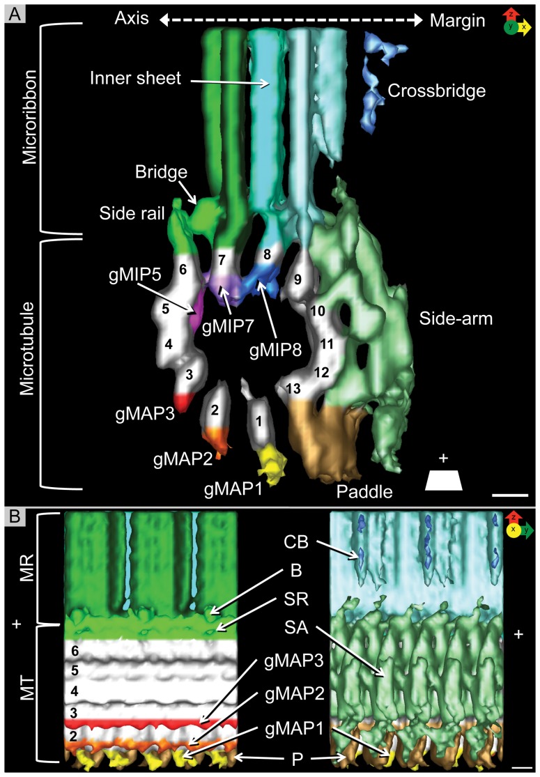
Figure 5. Isosurface representation of the grand average.
A) View along the microtubule axis torward the plus-end. Each protofilament is numbered clockwise, starting with 1 at the location of the seam (see Figure 4D). Microribbons consist of three parallel sheets: axis-facing sheet, inner sheet, and margin-facing sheet. The crossbridges are visible on the margin-facing sheet. The axis-facing sheet is connected to the side rail, a fibrous structure attached to protofilament 6, via the bridge. There are several giardial microtubule inner proteins (gMIPs) associated with the inner wall of the microtubule on protofilaments 5, 7, and 8 (gMIP5, gMIP7, and gMIP8). There are also giardial microtubule-associated proteins (gMAPs) attached to the outer wall at protofilaments 1, 2, and 3 (gMAP1, gMAP2, and gMAP3). The side-arms (SA) span protofilaments 9–12 and are associated with the paddle (P), which is connected to protofilament 13. B) Axis-facing (left) and margin-facing (right) views. The axis-facing side has 2 “naked” protofilaments (PF4 and PF5). All three gMAPs (gMAP1, gMAP2, gMAP3) are visible as well as part of the paddle (P). The side rail (SR) on protofilament 6 is connected to the axis-facing sheet of the microribbon via the bridge (B). On the margin-facing side, gMAP1 is barely visible behind the paddle. The side-arm covers the rest of the microtubule. The crossbridges (CB) are evident on the margin-facing sheet (M) of the microribbon. Scale bars = 5 nm.

