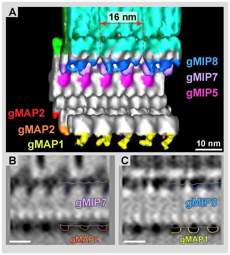
Figure 6. MIPs on the microtubule inner side of the grand average.
A) Isosurface representation of the inner microtubule wall and associated gMIPs. While gMIP5 and gMIP7 appear regularly every 8 nm, according to the αβ-tubulin dimer repeat, gMIP8 clearly exhibits a 16 nm repeat that reaches over two consecutive dimers along protofilament 8. Panels B and C show vertical 0.776 nm slices through the volume in A, cutting through protofilament 7 (B) and protofilament 8 (C), respectively. Scale bars = 10 nm.

