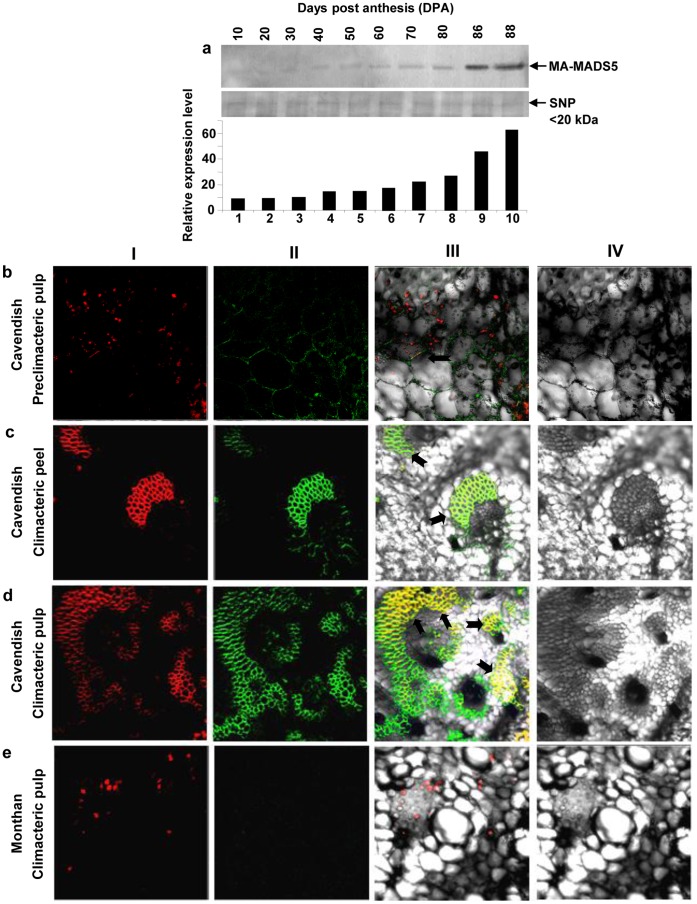
Figure 5. Immunoblotting and immunolocalization of MA-MADS5 protein in fruit tissues of banana.
a Changes of protein level of MA-MADS5 in developing fruit tissues were analyzed by immunoblotting using affinity purified anti-MA-MADS5 polyclonal antibody (1∶1000 dilution) (upper panel). Equal amounts of small nuclear protein (SNP) was loaded in each lane and shown as loading control (middle panel). Quantification of the data in (a) by densitometry (Bio-Rad Immage Densitometer G700) (lower panel). Representative images from at least three independent experiments are shown for Figure a. b–d In situ localization of MA-MADS5 protein using FITC-coupled affinity purified anti-MA-MADS5 IgG in the preclimacteric pulp (0 DAH), climacteric peel and climacteric (88 DAH) pulp tissues of Cavendish and in climacteric pulp tissues of Monthan. Nuclei were stained with DAPI (red fluorescence) (First row, I). MA-MADS5 was detected using anti-MA-MADS5 IgG coupled to FITC (green images) (Second row, II). Nuclear localization of MA-MADS5 (yellow fluorescence) has been shown in the merged pictures of I and II of b to e respectively (third row, III). Fourth rows (IV) of b–e indicate difference interference contrast pictures.

