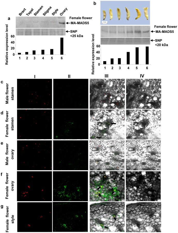
Figure 10. Immunodetection and in situ localization of MA-MADS5 protein in different floral tissues of banana.
a Changes in the protein level of MA-MADS5 in different tissues of female flower (upper panel). Equal amount of small nuclear protein (SNP) lane was loaded in each lane and has been shown as loading control (middle panel). Protein abundance in each lane in (a) was detected by densitometry (lower panel). b Various stages of development of banana female flower (first panel). Changes in the accumulation levels of MA-MADS5 protein in different stages of developing female flower ovary tissues (second panel). Equal amount of small nuclear protein lane was loaded in lane and has been shown as loading control (third panel). Quantification of the data in the second panel by densitometry (lower panel). c–g Immunolocalization of MA-MADS5 protein in different tissues of banana flower including male flower stamen, female flower stamen, male flower ovary, female flower ovary and female flower style respectively. The legends of the panels I–IV for c-g were identical to those described in Figure 5b–e. Representative images from at least three independent experiments are shown.

