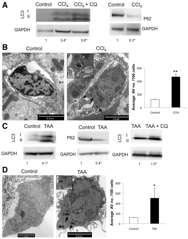
Figure 1.
Autophagy is up-regulated in stellate cells after liver injury in vivo. (A and C) Immunoblots of stellate cells isolated from wild-type mice after acute liver injury induction with (A) CCl4 and (C) TAA with and without addition of CQ showing an increase of LC3II conversion and decrease in P62. (B and D) Electron micrographs of whole liver tissue, showing stellate cells after (B) CCl4 and (D) TAA treatment. Arrows indicate AV. (Right) Electron micrograph quantification of AV number per 100 cells (★P < .05, ★★P < .001; error bars indicate SEM). Protein ratios (normalized to GAPDH) were used to quantify fold change relative to control and are shown below each blot. Data represent the mean value of at least 3 experiments (★P < .05), and 3 animals per condition were used in this experiment.

