Figure 3.
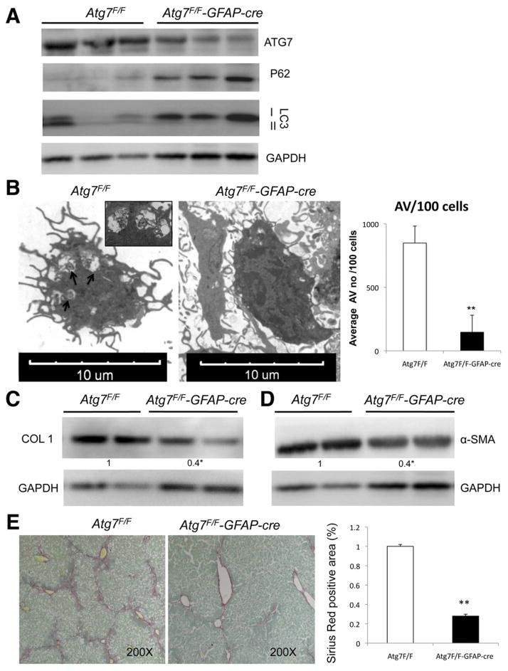
Autophagy regulates stellate cell activation and fibrosis in vivo. (A) Immunoblots of stellate cells protein isolated from Atg7F/F and Atg7F/F-GFAP-cre mice showing decreased expression of ATG7, LC3-II, and increased P62. (B) Electron micrographs of stellate cells isolated from Atg7F/F and Atg7F/F-GFAP-cre mice and AV quantification (right) depict a significant decrease of AV in transgenic animals. Arrows indicate AV. (C and D) Immunoblots for (C) collagen type I and α-SMA in isolated stellate cells from Atg7F/F and Atg7F/F-GFAP-cre mice after chronic liver injury with CCl4. (E) Whole liver sections after chronic liver injury with CCl4 were stained for Sirius Red. (Right) Quantification of Sirius Red–positive area. ★P < .05, ★★P < .001. Error bars indicate SEM. Protein ratios (normalized to GAPDH) were used to quantify fold change relative to control and are shown below each blot. Data represent the mean value of at least 3 experiments (★P < .05) and a total of 18 animals: 9 Atg7F/F and 9 Atg7F/F-GFAP-cre.

