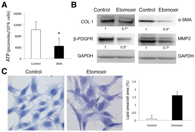
Figure 5.
β-oxidation regulates stellate cell activation. (A) Autophagy was inhibited in JS1 cells with 3-MA, and ATP content was determined at 12 hours after treatment. (B and C) β-oxidation was blocked with etomoxir in JS1 cells after 12-hour expression of COL1, α-SMA, and β-PDGFR was determined by (B) immunoblot analysis and (C) LD content was examined by ORO staining (right, quantification of ORO-stained area). Levels of ATP are expressed in picomoles per 106 cells. Error bars indicate SEM. Protein ratios (normalized to GAPDH or tubulin) were used to quantify fold change relative to control and are shown below each blot. Data represent the mean value of at least 3 experiments (★P < .05).

