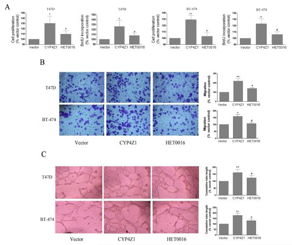
Fig. 3.
The conditioned medium from CYP4Z1-expressing T47D and BT-474 cells enhanced proliferation, migration and tube formation of human umbilical vein endothelial cells (HUVECs). (A) Cell proliferation. HUVECs were plated at 3×10³ per well in 96-well culture plates, and then treated with the conditioned medium as in the presently described method. Cell proliferation was measured using the MTS and BrdU incorporation assays. (B) Migration assays. HUVEC migration assays were performed using culture supernatants that were derived from T47D or BT-474 cells transfected or untransfected with CYP4Z1, and cell migration was measured in a 16-h Transwell assay in a blinded manner. (C) Tube formation assays. Cells were seeded on matrigel-coated wells in the presence of different conditioned media, as indicated, and incubated for 18 h to form a capillary network. The total number of branched tubes was then counted. Results are shown as mean ± S.D. from 3 independent experiments (n=3). *P < 0.05, **P < 0.01 vs. vector control group; #P < 0.05 vs. T47D-CYP4Z1 group.

