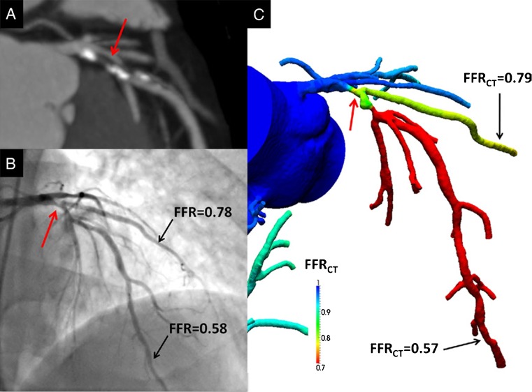
Fig. 1.
Anatomically obstructive stenosis with a lesion causing ischaemia (a) Multiplanar reformat of coronary computed tomography angiography demonstrating obstructive (> 50 %) stenosis (white arrow) in the proximal portion of the left anterior descending (LAD) artery. (b) Invasive coronary angiography confirms the LAD stenosis (red arrow) with corresponding haemodynamically significant reductions in coronary pressure in the first diagonal branch (0.78) and distal LAD (0.58) by FFR. (c) Noninvasive computation of FFR from FFRCT of the first diagonal branch (0.79) and distal LAD (0.57), demonstrating lesion-specific ischaemia of the proximal LAD stenosis. (Reproduced with permission from Koo et al. J Am Coll Cardiol. 2011 [36])

