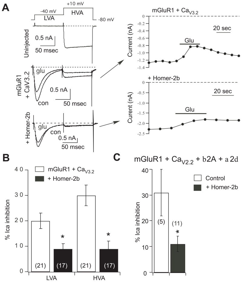
Fig. 4.
Overexpression of other voltage dependent calcium channels in SCG neurons does not preserve coupling to mGluR1 in the presence of Homer-2b. A, sample current traces (left) and time courses (right) illustrating the two-step voltage protocol (top) used to separate low voltage activated (CaV3.2) currents (LVA) from the native high voltage activated currents (HVA), and inhibition by 100 μM glutamate. Uninjected SCG neurons (upper left) showed no LVA currents. Neurons injected with mGluR1 and CaV3.2 (middle left and upper right) had prominent LVA and HVA currents, both of which were modulated by 100 μM glutamate (Glu). Additional co-expression of Homer-2b (lower left and lower right) uncoupled both the LVA and HVA currents from glutamate modulation. B, Average (± SEM) LVA and HVA calcium current inhibition by 100 μM glutamate in cells with the indicated expression. Number of cells is shown in parentheses. C, Average (± SEM) calcium current inhibition by 100 μM glutamate in control cells (expressing mGluR1, CaV2.2, and accessory proteins) and in cells co-expressing Homer-2b. Asterisk indicates a statistically significant difference from paired control (without Homer-2b; p ≤ 0.05). Number of cells is shown in parentheses.

