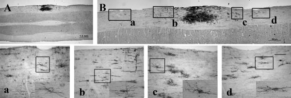
Fig. 4.
Migration of NPCs from transplantation site along the spinal white matter. AP staining shows NPCs grafted into spinal white matter (A) migrating rostrally and caudally. Panels A and B show the entire NPC graft together with the migrating cells. Panels a–d show higher magnification of images from different regions along the migration path of the AP-positive cells corresponding to the labeled boxes in B. The insets shown in panels a–d demonstrate the morphology of the migrating AP cells. Scale bar = 1 mm in A; 500 μm in B; and 100 μm in a–d. Rostral at the left side in Figure 4A and B.

